The Franco-Indian Workshop on Microscopy, held from 6 – 10 October 2025 at Ashoka University, brought together researchers from India and France to explore cutting-edge imaging in disease biology. The five-day event fostered collaborations, hands-on microscopy learning, and vibrant scientific dialogue across borders.
When I look back at the five days of the Franco-Indian Workshop on Microscopy 2025, held from 6 – 10 October at Ashoka University, I recall not only the hum of scientific conversations but also the quiet joy of watching months of planning unfold into an event alive with connections and collaborations.
This workshop went far beyond a typical academic gathering. It marked a landmark initiative under the Franco-Indian Campus in the field of Life Sciences for Health, a virtual consortium connecting leading scientists and institutions across India and France. Nearly 20 French researchers from institutions such as Université Côte d’Azur, Sorbonne Université, and École normale supérieure de Lyon, alongside Indian researchers from Indraprastha Institute of Information Technology (IIIT) Delhi, Indian Institute of Technology (IIT) Delhi, Indian Institute of Science (IISc), National Centre for Biological Sciences (NCBS), and the Indian Institutes of Science Education and Research (IISER) Kolkata and Thiruvananthapuram, participated in the workshop. For me, it was also a deeply personal project, one I guided from the first email thread to the final group photograph.
We set out with an ambitious goal: to create a space where biologists from both countries could connect over a unifying theme — microscopy in disease biology. Kasturi Mitra and Sandeep Ameta, organisers from Ashoka University, thoughtfully curated the scientific programme and shaped the structure of the research talks and discussions across the two days of the meeting. I, along with Anupama Ambika Anilkumar from Ashoka Global Research Alliances, coordinated across time zones, institutions, and expectations, aligning every detail — from designing the schedule, curating sessions to managing travel logistics. When the first participants arrived on campus, I realised that every late-night call and checklist had been worth it.
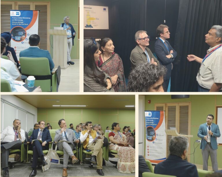
Inauguration by French delegation on Day 1 of the workshop. Photo Credit: Ashoka University. Collage by Ankita Rathore.
The conference opened on 6 October 2025 with the theme “Disease Biology: Bench to Bed and Back.” Grégor Trumel, Counsellor for Education, Science & Culture and Director, French Institute in India; Tonmoy Kundu, Head of Global Sales, Zeiss Microscopy; Nitin Seth, Director, Indo-French Centre for the Promotion of Advanced Research (CEFIPRA); and Gilles Alcaraz, Director, CNRS India, inaugurated the event and set an inspiring tone. Their remarks reflected a shared optimism about the strengthening Indo-French scientific partnership. Trumel noted,
This workshop stands as a testament to how far a collaboration can go, with India and France advancing science for the greater good.”
K. VijayRaghavan, Science Advisory Chair; Anurag Agrawal, Dean, Trivedi School of Biosciences; and Gautam Menon, Dean of Research, all from Ashoka University, welcomed the delegates. As I listened to them highlight Ashoka’s expanding role as a hub for interdisciplinary research, I felt reaffirmed about the purpose that had guided every planning step of the workshop.
The research talks across the two conference days covered a fascinating range of themes: from cell morphogenesis and cancer biology to neural imaging and artificial intelligence in microscopy. In the keynote lecture, Maithreyi Narasimha, TIFR Mumbai, showed how imaging continues to redefine our understanding of dynamic biological processes and reminded us that seeing truly deepens understanding.
Matteo Rauzi, Université Côte d’Azur, opened the scientific sessions with a striking demonstration of multi-view lightsheet microscopy and laser manipulation to study tissue folding during embryonic development. L.S. Shashidhara, NCBS, offered a translational perspective through molecular and cellular profiling of triple-negative breast cancer in Indian cohorts, showing how imaging aids context-specific disease understanding. Muriel Grammont, ENS Lyon, examined how mechanical and genetic factors drive cell shape changes during epithelial transitions. Pratik Kumar, NCBS, discussed how organic dyes can function as molecular tools beyond imaging, expanding their applications in probing biological systems.
Nicolas Heck, Sorbonne Université, presented 3D confocal imaging and analysis to study connectivity changes in the rodent brain, linking cellular imaging with neuroscience. Tavpritesh Sethi, IIIT Delhi, then highlighted how transformer-based artificial-intelligence architectures, such as Vision Transformers, can transform histopathology by enabling accurate and interpretable analysis of whole-slide images for disease diagnostics.
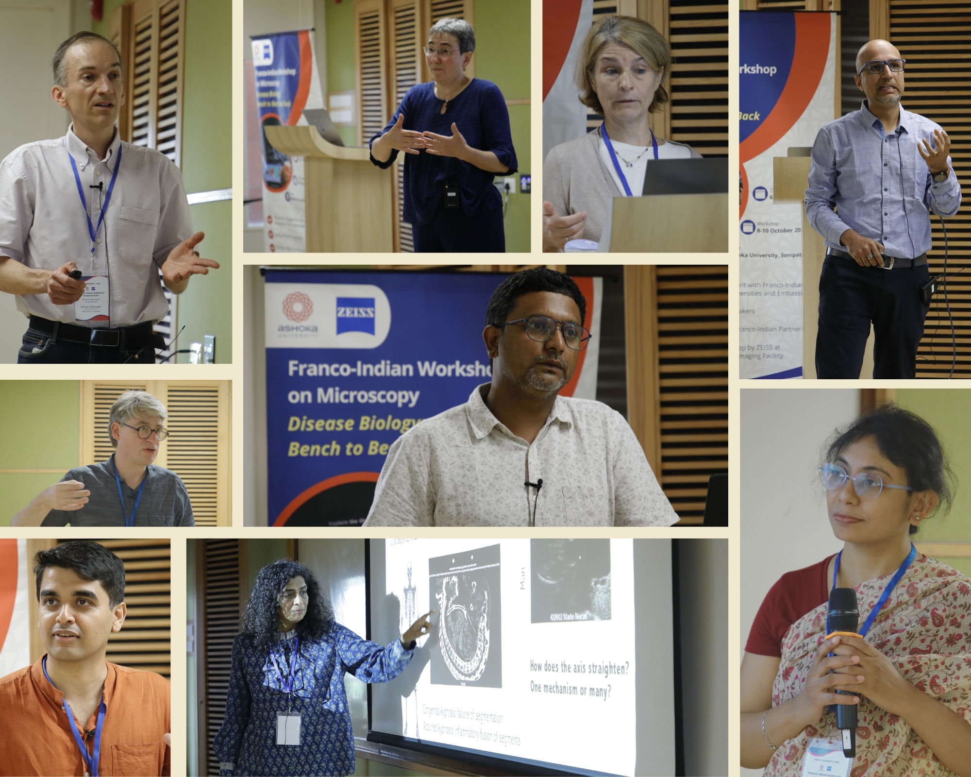 Speakers over 2 days of the workshop. Photo Credit: Ashoka University. Collage by Ankita Rathore.
The second day sustained this momentum with equally engaging sessions. Anup Padmanabhan, Ashoka University, explored the mechanobiology of host – pathogen interactions, while Kasturi Mitra, Ashoka University, revealed how redox-tuned mitochondrial networks prime stemness through quantitative microscopy. Mireille Cormont, Université Côte d’Azur, along with Saravanan Palani and Shovamayee Maharana from IISc, expanded the discussion to cytoskeletal imaging, adipose tissue inflammation, and phase separation biology. Sandeep Ameta, Ashoka University, examined the dynamics within liquid – liquid phase-separated droplets, while Deepak Nair, IISc, and Sylvain Feliciangeli, Université Côte d’Azur, showcased how nanoscale organisation governs ion channel and synaptic function in the brain.
Participants also engaged in parallel breakout sessions exploring microscopy in fundamental and translational biology, high-throughput imaging, and image analysis approaches. Collectively, these sessions showed how microscopy today extends far beyond imaging, it drives discovery, integration, and dialogue across biological scales, from molecules to whole organisms.
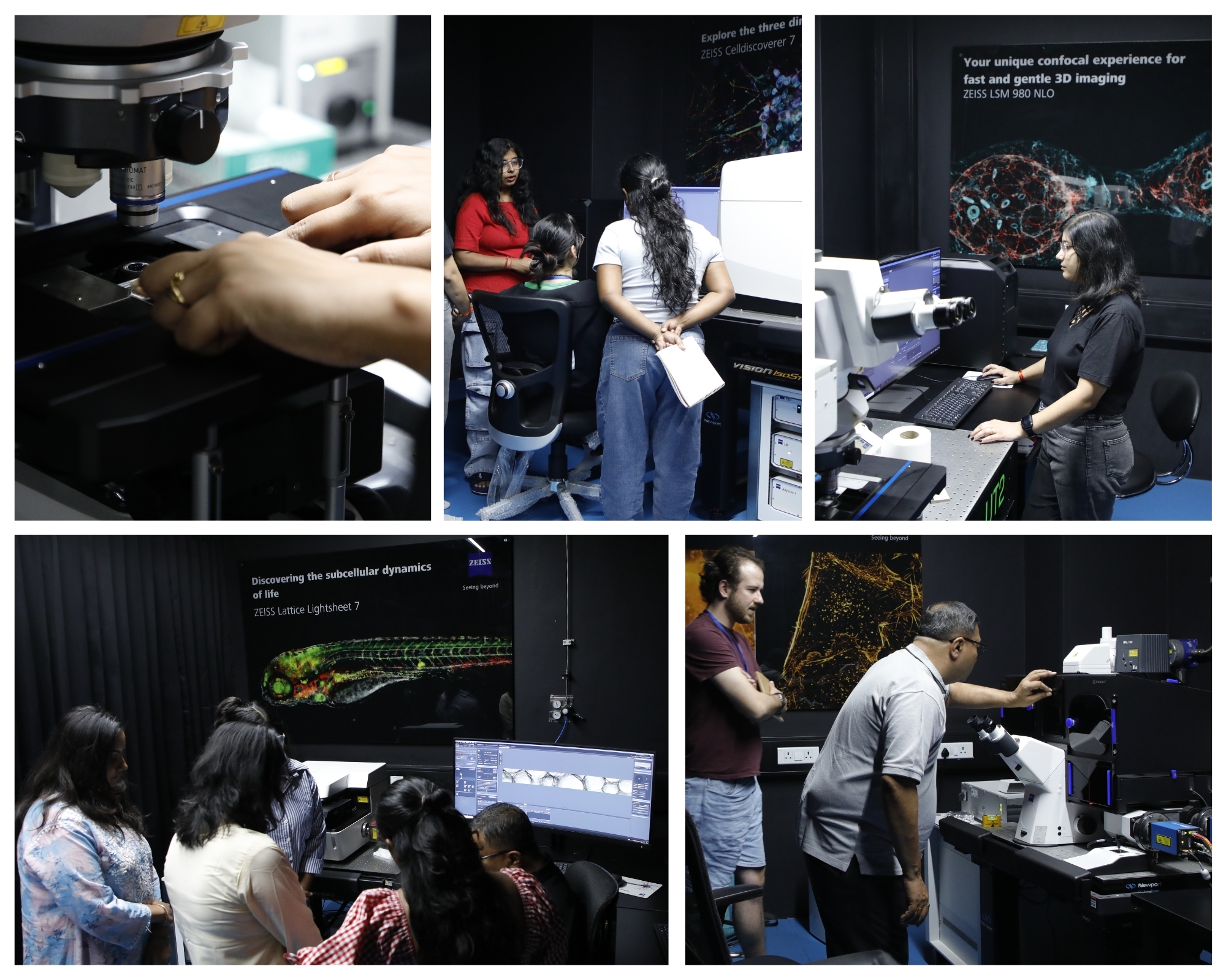 Glimpses from the three-day hands-on workshop at the Ashoka-Zeiss Core Imaging Facility. Photo Credit: Ashoka University. Collage by Ankita Rathore.
The two-day meeting concluded with a visit to the Ashoka – Zeiss Core Imaging Facility, a collaborative platform supporting advanced imaging research, underscoring the shared commitment of France and India to foster excellence in life sciences research. We organised a hands-on microscopy workshop, from 8 – 10 October 2025, led by Rishi Kant from Zeiss Microscopy for 16 selected participants from Franco-Indian Campus partner institutions, including Université Côte d’Azur, Sorbonne Université, IIT Delhi, IIIT Delhi, Vellore Institute of Technology, and IISER Kolkata. Participants brought their own microscopy samples and trained on advanced imaging systems, exploring high-resolution microscopy techniques.
As one of the organisers, I experienced the other side of the story, the one that rarely appears in conference reports. The weeks leading up to the event overflowed with spreadsheets, video calls, and coordination marathons. I arranged travel for nearly 60 participants, managed logistics for international guests, and refined every detail of the scientific programme. Behind every “successful scientific meeting” stands a web of collaboration, patience, and trust. Our organising team ensured that participants could focus on science while we quietly handled everything else in the background.
The workshop did more than bring together researchers from two nations; it strengthened a network, inspired collaborations, and reinforced the role of microscopy as a bridge between basic and applied science. It reminded me why such platforms matter: they build not only research partnerships but also communities founded on trust, dialogue, and mutual respect.
We owe deep gratitude to our collaborators — CEFIPRA, CNRS India, the French Institute in India, and Zeiss Microscopy — for their unwavering support. Their belief in sustained Indo-French engagement in life sciences made this Franco-Indian Workshop on Microscopy at Ashoka University possible.
Images are for reference only.Images and contents gathered automatic from google or 3rd party sources.All rights on the images and contents are with their legal original owners.