Our Terms & Conditions | Our Privacy Policy
Chinese scientists use ‘ionic glass’ to make organs transparent without slicing, unlocking 3D ‘X-ray vision’ of the human body
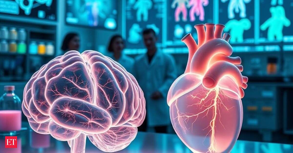
Chinese researchers have developed a pioneering method that turns organs such as the brain and heart transparent without damaging their structure, a breakthrough that could transform medical imaging and disease research.
Scientists believe that the ionic glass method may allow them to visualize entire organs in three dimensions, overcoming long-standing barriers in the study of complex tissues such as neurons, blood vessels, and cardiac networks.
Traditionally, to study something like the brain or heart in detail, scientists had to dissect the organ into slices and then examine them under a microscope. This destroys the intact structure and makes it harder to understand how all the parts are connected in 3D.
The new “ionic glass” method makes the organ transparent while keeping its original shape intact.
Why transparency matters
Live Events
Scientists have tried to peek inside whole organs without cutting them apart. But biological tissues are naturally opaque and block light, while fluorescent dyes, used to highlight cells and molecules, don’t penetrate easily.
Existing “clearing” methods can make tissue transparent but often distort the samples, causing organs to expand, shrink, or lose delicate details.
Frozen methods risk ice crystal damage. This has limited researchers’ ability to study organs intact.
The ‘ionic glass’ solution
The Chinese team, led by Tsinghua University and including researchers from Beijing Chaoyang Hospital, Shanxi Medical University, Beijing Qingzhun Medical Technology Company, and Fudan University, turned to ionic liquids, special solvents that stay liquid below water’s boiling point.
When organs are treated with these liquids, they enter an “ionic glassy state.” In this state:
- Tissues become transparent while preserving their original shape and microstructures.
- Samples can be stored cold for long periods without ice damage.
- Fluorescent dyes glow 2–30 times brighter, making even faint biological signals visible.
What the researchers found
Using this method, the team successfully examined the micro-connectivity of human neurons and even identified subtle differences in impulse control compared to non-human brains.
Because the process enhances visibility of rare proteins and neural pathways, it could help create highly accurate 3D maps of entire organs, something researchers have long sought but struggled to achieve.
Potential applications
According to the South China Morning Post, the university said the advance could be a game-changer, and they would work on precision medicine and intelligent diagnostics with it.
The Tsinghua University’s social media page described their technique as providing “X-ray vision for tissues,” combined with a built-in system to manage sample preparation, dye staining, and 3D reconstruction.
Images are for reference only.Images and contents gathered automatic from google or 3rd party sources.All rights on the images and contents are with their legal original owners.
Aggregated From –
Comments are closed.