Our Terms & Conditions | Our Privacy Policy
Subscribe now for full access and no adverts
We have covered the growing area of palaeoproteomics – the study of proteins in archaeological remains – a few times in ‘Science Notes’ (see, in particular, CA 338 and 353). The research up to this point has been largely focused on the analysis of hard tissues (that is, bones and teeth) with research on ancient soft tissues primarily conducted on animal skins (see CA 381). But, since 10% of human proteins are found in bone and around 75% in internal organs, we are missing a vast array of information by ignoring human soft tissue. While hard tissue is invariably more commonly preserved, and thus has rightly been the focus of investigation, the preservation of soft tissue is not unheard of, particularly in the waterlogged contexts that are so common across the United Kingdom and Ireland. This month, we will examine pioneering new research into how these preserved soft tissues may be assessed using proteomic techniques.
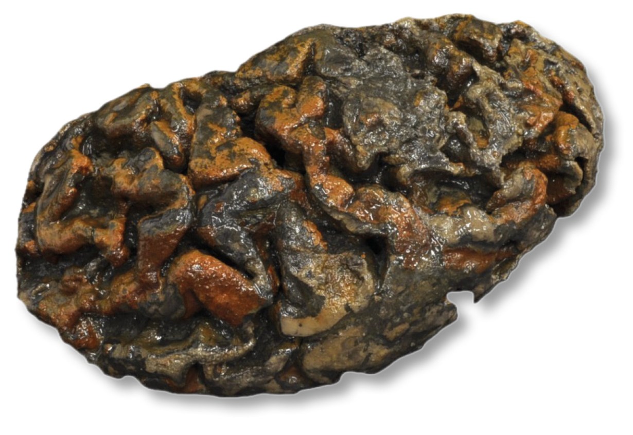
A team from the University of Oxford, led by Alexandra Morton-Hayward, recently developed a method for successfully extracting and identifying proteins from ancient internal organs. This was not a straightforward task, primarily because it is difficult to gain permission to do destructive sampling on soft tissue from human remains. The excavation of the former Blackberry Hill Hospital in the Fishponds area of Bristol, undertaken in 2018-2022, provided a great opportunity to rectify that, however, as around one in ten of the individuals buried there had evidence of preserved brain tissues (456 out of approximately 4,500). While this site had had many iterations, first as a prisoner-of-war camp, then as a hospital, workhouse, and back to a hospital again, most of the burials were from when it functioned as Stapleton Workhouse between 1837 and 1890 (CA 418).
Once a sample had been obtained, the next step was to figure out how to break down the lipid bilayers that form the cellular membranes in order to release the proteins within. Testing out ten different methods, the team found that urea (a key component of urine) was the most effective at this, with techniques that used urea-lysis before mass spectrometry producing the broadest range of peptides. The team argue that this is probably due to the fact that urea breaks down non-covalent interactions, such as hydrogen bonds, so that proteins become less hydrophobic – meaning that they are more likely to become unfolded in a solution, and can be successfully extracted.
Following the extraction of the proteins through lysis, the researchers needed to establish the best way to separate and identify them. The usual method is to separate them using liquid chromatography and then identify them using mass spectrometry. The team found, however, that liquid chromatography-mass spectrometry (LC-MS) was more effective when coupled with a method called high-field asymmetric-waveform ion mobility spectrometry (FAIMS), which separates ions based on how they move in an electric field. By using LC-FAIMS-MS, they were able to identify over 1,200 proteins, which was 40% more than with just LC-MS alone. Importantly, they also found that, although they started with 50mg of brain matter, only 2.5mg were needed for successful extraction and identification, indicating that very small amounts of tissue will be sufficient for future analysis.
Despite the success of this study, however, there are still limitations. Only a fraction of an organism’s proteome is expected to survive archaeologically, and those that persist are likely to be those that are more abundant and/or have specific structural features that allow them to survive. As such, there will be biases in the types of proteins that are identified from a sample. For example, while only 13% of a brain’s proteome is hydrophobic (meaning it repels water), on average 39.6% of all identified peptides had this quality. This suggests that either the techniques used preferential identified hydrophobic proteins, or that these proteins are more likely to be preserved archaeologically. The researchers suggest that it is probably the latter as, when soft tissue decays, the breakdown of proteins produces acidic byproducts such as lactic acid, fatty acids, and negatively charged amino acids. These acids lower the pH of the surrounding tissue, meaning that hydrophobic proteins are more likely to persist than hydrophilic ones, as they tend to thrive in all but the most alkaline environments.
Still, these results represent the largest and most diverse palaeoproteome ever reported, and are a testament to the potential that future research in this area holds of being able to inform more deeply our knowledge of human health and well-being in the past.
The full results of the study were recently published in PLOS One: (open access).
Text: Kathryn Krakowka / Image: Alexandra Morton-Hayward
Images are for reference only.Images and contents gathered automatic from google or 3rd party sources.All rights on the images and contents are with their legal original owners.
Aggregated From –
Comments are closed.